
Putting names to the waves:

As you can see above, each wave or deflection is represented by a letter. Not all waves are always present. There are other wave forms from different pathologies that will be mentioned later in the tutorial.
Quick refresherDepolarization - Electrical dischargeRepolarization - Electrical recharge
P wave - The P wave is normally the first sign of electrical output. This represents atrial depolarization. Usually the SA node, but may represent any form of mono-focal atrial depolarization. The P wave is usually less than 120 ms (0.12 sec), or 3 small boxes. The amplitude should be less than 0.25 mv.
Atrial repolarization is thaught to be hidden inside the QRS complex, and it doesn't give off enough energy to show on an ECG. A more complex theory is that atrial repolarization occurs with the downslope of the P wave. This theory is due to the fact that atrial repolarization should occur much faster than it would if it were buried within the QRS complex.
QRS Complex - Not always encompassing all 3 parts and sometimes more than one R wave. The QRS complex represents ventricular depolarization. The QRS complex is usually between 40 ms and 120 ms (0.04 sec - 0.12 sec), or 1 to 3 small boxes.
The image below shows the different examples of QRS complexes. The apostrophe stands for prime.

Q wave - The first negative deflection on a normal ECG in most leads. The Q wave may or may not be present as a normal physiological variant. This represents the initial faze of depolarization of the ventricular myocardium; depolarization of the interventricular septum.
R wave - Normally the most prominent positive deflection on an ECG. Represents early ventricular depolarization.
S wave - The negative deflection following the R wave. This represents late ventricular depolarization.

T wave - The T wave is normally the next positive deflection following the QRS complex. Represents ventricular repolarization.
U wave - Often unseen or hidden in the T wave. Thought to represent repolarization of the perkinje fibers.
As we start to learn about different ECG interpretations and pathologies, you will see how important intervals and measurements are. Each wave, and interval has a normal amplitude and time measurement, and variation may be severe. Take a look below:

PR Interval - From the very beginning f the P wave to the first deflection of the QRS complex. The PR Interval should end on the isoelectric line. The normal PR interval last from 120 ms to 200 ms (0.12-0.20 sec).

PR Segment - The isoelectric line between the end of the P wave and the beginning of the QRS complex. This pause in electrical activity depicts the pause of the impulse at the AV node.
QT Interval - From the beginning of the QRS complex to the end of the T wave. These intervals are normally shorter in men than in women. For more on QT intervals and possible pathologies click here.
ST Segment - The very end of the S wave, where it meets the isoelectric line, is known as the J point. This point may be above or below the isoelectric line depending on certain pathologies. The ST segment begins at the J point and ends at the very beginning of the T wave.

RR Interval - R to R interval. Measured by the blue line in the image above. The RR interval should remain consistent. This is a measure of the regularity of ventricular depolarization. Below is an example of a regular RR interval.

PP Interval - P to P interval. Measured by the yellow line above. The PP interval should remain consistent as well. This is a measure of the pacemaker's regularity. Below shows how the PP interval should march out.

It is important to learn the normals before we get to all the arrhythmias you want to learn.
Arrhythmia - Absence of rhythm. Anything other than normal sinus rhythm.
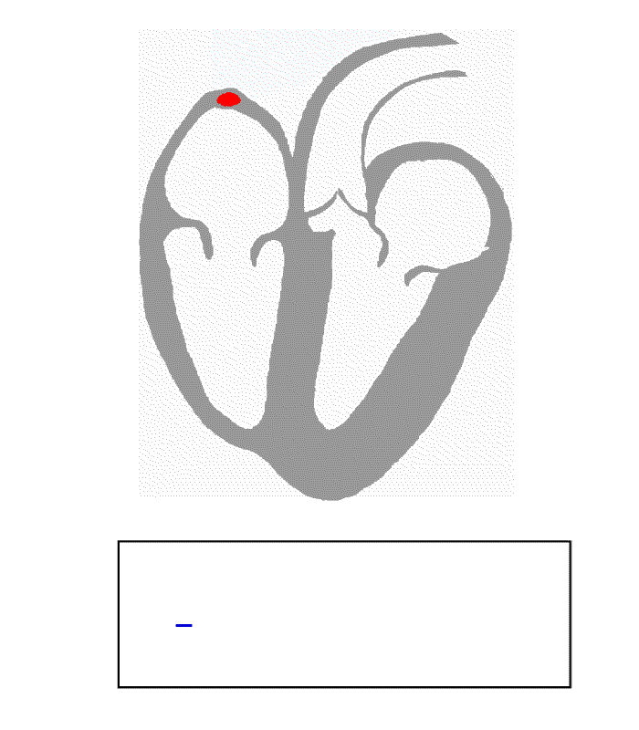
Blood pours into the right atrium from the vena cava and the left atrium from the pulmonary vein.The SA node fires a signal representing a P wave on the ECG. A this point the atria contract, ejecting their blood into the ventricles.As the impulse travels to the AV node, the PR segment shows the pause in electrical activity. This pause allows the ventricles to fill to capacity.The interventricular septum then depolarizes, and then the rest of the ventricular myocardium. We see this as a QRS complex.The blood ejects out of the ventricles. The Ventricular myocardium then recharges which is displayed as a T wave.


1 comment:
finally, after 8 years of self learning ecg, i have related the electrical impulses, ecg tracing and myocardial contractions. thanks!
Post a Comment