Click here for part 1.
Click here for the next part.
Now that we know how the heart functions mechanically, it is time to learn what initiates that functioning. The following is a brief overview of cardiac electrophysiology.
Automaticity - In cardiology, the ability of an individual myocardial cell to depolarize spontaneously.
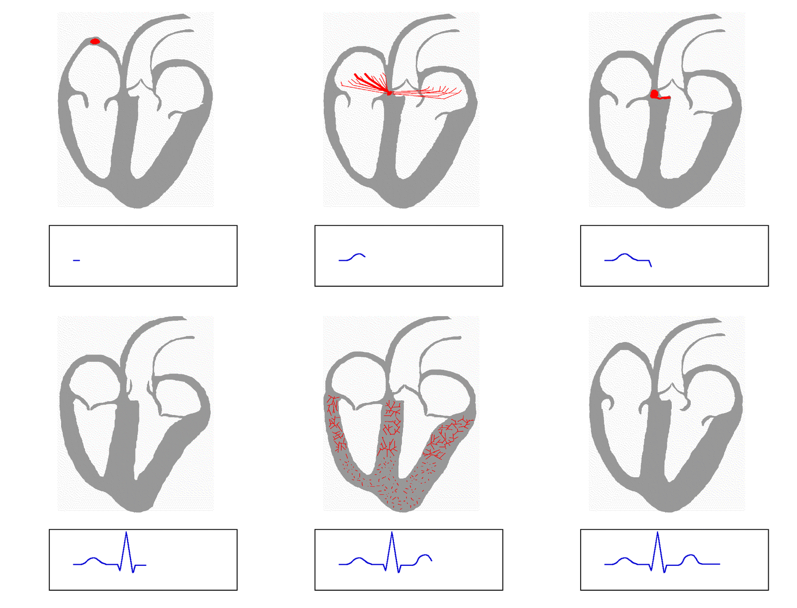
The myocardium consists of special pathways of conduction. This conduction causes subsequent contractions when working properly. The system is an integral part of healthy cardiac functioning. It is important to remember that the two atria contract almost simultaneously, and then shortly there after the two ventricles do the same thing. So imagine this as top-bottom-top-bottom. Without one set of depolarization, the body suffers greatly.
The sinoatrial node (SA node) - The SA node is the heart's physiological pacemaker. It is located in the right atrium, roughly in the top right corner. The SA node has an intrinsic rate of about 60 to 100 beats per minute (bpm). In normal conduction, all cardiac impulses originate at the SA node. Impulses travel from the SA node and right atrium across the inter-atrial septum to the left atrium. This process initiates atrial depolarization, which causes atrial contraction and the subsequent exchange of blood from the atria to the ventricles.
The atrioventricular junction (AV junction) - This is a term used to describe the next to structures, the AV node and the bundle of his. This is a term used mostly when speaking about the electrocardiogram. Certain arrhythmias may be attributed to the AV junction, because it is impossible to know exactly where the impulse is originating.
The atrioventricular node (AV node) - Named due to its placement, as the conduction link between the right atrium and ventricle; the AV node is the next stop for cardiac conduction. It is located just superior to the atrioventricular septum. The AV node is a yield sign, so-to-speak. It slows the conduction from the atria to the ventricles to prevent overlap in depolarization. This in turn allows for adequate ventricular filling, and Frank Starling's mechanism to work. The intrinsic firing rate for the AV node is 40 to 60 bpm.
It is important that the intrinsic rate of each structure is slower the further you go down the conduction system. This helps prevent an unwanted takeover of the heart's pacemaker. As you will learn, certain conditions bypass this security system.
The bundle of his - Located in the ventricular septum. The bundle of his is comprised of fibrous tissue. It is the first part of the conduction chain below the AV node.
The bundle branches - The bundle of his branches off into two main bands of conduction. These are known as the bundle branches. The right and left bundle branches facilitate further ventricular conduction. The right bundle branch has a single fascicle, while the left bundle branch consists of three fascicles; although it is common to state that the left bundle brach only has two fascicles. The left anterior, left posterior, and septal fascicles are all located on the left bundle branch.
The purkinje fibers - These are located just below the endocardium throughout the ventricular walls. They are the conduction pathways of the ventricles. The intrinsic rate for the purkinje system is about 20 to 40 bpm.
Depolarization - The firing of the cardiac cells, initiating or continuing an impulse.
Repolarization - The regenerating of the cardiac cells. When the cell is not firing, but building up the energy to depolarize again.
So to sum things up the SA node initiates an impulse which travels across both atria causing atrial depolarization. This conduction causes the atria to contract and eject it's blood into the ventricles. The AV node pauses conduction momentarily while the ventricles fill. The atria then repolarize while the ventricles depolarize. The ventricles depolarize when the conducted impulse travels down the bundle of his, bundle branches, and purkinje fibers. This depolarization causes the ventricles to contract and eject it's blood. The ventricles then repolarize and the cycle begins again shortly after.
There are many pathologies that distort the normal flow of cardiac conduction. These will be described as I describe the ECG interpretation for each.
The electrocardiogram (ECG) is a transthoracic interpretation of the heart's electrical activity over time.
I am not going to get in to action potentials and electrolyte exchange just yet. This is just the basics. The next post in this series will begin our ECG tutorial.


No comments:
Post a Comment