The Electrocardiogram - Part V
Adam Thompson, EMT-P | 11:09 PM | Cardiology, Education | 0 comments
Strip Tease 14: Answer
Adam Thompson, EMT-P | 11:05 PM | ECG/EKG Archive | 4 comments
Addition by Rogue Medic 00:17 9/18/09.
My little bit of strip marking to show what I was looking at with this strip. These are just different ways of coming to the same conclusion. In EMS there is rarely just one right way of doing things. The response of the patient is the best indicator of what is right. Sometimes nothing is right.

.
Sick Kid
Shaggy | 9:26 PM | Clinical Discussion, Diabetes | 14 comments
My goal with this 3 part post is to discuss a particular disease state focusing on the pathophysiology and the current treatment guidelines and their rationale. Let's start with the presentation:
Introduction
You are the medic working with with an EMT on a 12 hour shift in an urban low income, mixed race neighborhood. You have been running non-stop for 9 hours, doing two BLS and 5 ALS trips, and you are hungry, tired, and have 4 PCRs to type up yet (while your partner only has two). It is a typical cold winter day with snow flurries. The heater is on to not only warm you guys but as a half hearted attempt to keep your food warm that is sitting on the dash over the defroster duct. You have been nibbling at your sandwich over the past couple of hours. Just as you take a bite, you get a call for a 9 YOM "sick and vomiting". Your partner slams her hand on the steering wheel while you yell out a few expletives. "This kid better be sick!" Your partner yells out, "Who calls an ambulance for vomiting?" Why ask? We get these calls everyday, and will continue for a long time.
You pull up to an old row house in this old mill town and see a somewhat neat front porch atop a flight of stairs with bikes in the front yard. You are already thinking you aren't carrying someone down these stairs for just feeling sick. This kid is going to walk. You grab the clipboard while your partner grabs the jumpbag and you ascend the stairs to the house. The door opens and an overweight but neat appearing woman in her late 20s opens the storm door. She appears concerned and tells you it is her son who is sick. She holds the door open as you and your partner pass through. She tells you he is upstairs in bed and scurries past you to lead the way to his room. Great. More steps. This kid is definitely walking.
Chief Complaint
As you climb the stairs, you try to illicit a history of present illness. She tells you he has been sick the past few days, progressively getting worse. "So why now are you calling?" You ask. She turns around to see the expression on your face as she senses a slight sarcasm in your voice. You quickly add, "I mean, what prompted you to call now? What is worse this evening?"
"He has been throwing up all day and now and is weak, laying in bed all day," she says as she turns the corner and heads down the hall. "He won't even play his video games."
"Has he had a recent fever?", you ask. She tells you she didn't notice and does not have a functioning thermometer. "Do you think it is the flu?" she asks. You shrug your shoulders. You ask about his medical history. He has none other than a T&A (tonsillectomy/Adenoidectomy) done last year.
Your partner asks, "Did you call your PCP for guidance?" and the woman answers that she thought she should just take her child to the ER. Yeah, the ER-the abused modern day clinic. Your partner asks if she has a car to drive him to the hospital and the woman says no. Of course not. If she needed a ride to Walmart she would have no problem getting one, but to the hospital, the ambulance is always free. Or so it seems to many. You keep those thoughts to your self. You are used to this.
General Impression
As you walk in the room, you see the thin child, of normal size for his age, laying in bed. He shares the room with his younger brother and it is expectedly messy. Though it is a boy's room, you can smell someone was painting nails or something similar. He makes eye contact with you but gives a half hearted smile, showing signs he is tired of being sick. Looking at him makes you overcome with pity. You have forgotten you are a health care provider, often called to replace the MD who did house calls in the past. Despite how busy you are, it is not this kid's fault he is sick. He did not plan this. Despite whether a patient needs transport or not, you have a responsibility to alleviate pain and suffering, and this kid appears to be suffering in some way. This family called you for help and you have an obligation to help them.
Assessment
You talk to the kid and he is alert and oriented but appears slightly pale and tachypneic. Actually, he appears to be hyperventilating at 28 and regular but he denies any SOB and you have seen much worse on healthier patients. His skin is warm and dry and he tells you he is thirsty and hungry but cannot keep anything down.
In the meantime, your partner get vitals:
P=110, S/R
BP=90/58, manually
R=28, deep and regular
T=37.6
Pulsox=97% RA
Your partner asks you if you want to make this ALS or BLS and how do you want to get him to the truck. What is your answer? What is your general impression of this patient? Are the vitals within normal limits for a 9 year old. Does he need to go the hospital or should you call his pediatrician about scheduling an urgent office visit the next day? Would you refer the child to bee seen at the local urgicare center or clinic in the morning? Are there any additional isolation precautions you should consider? What other assessment information would you like (or need) to guide your treatment plan for this patient?
Lee County EMS Airway Management Guidelines
Adam Thompson, EMT-P | 8:28 PM | Airway, Airway Management, Respiratory | 0 comments
CORE PRINCIPLE
AIRWAY, VENTILATION, AND OXYGENATION
AIRWAY ADEQUACY
IMPORTANT CONCEPTS IN AIRWAY MANAGEMENT
The assessment and management of a patient’s airway is the crucial initial priority in all circumstances. Usually, this is easily accomplished when faced with a talking, breathing, and coherent patient. Other times it is more difficult to determine if the patient’s airway is compromised, ventilatory rate inadequate, or air exchange is poor. Additionally, there may be circumstances when airway adequacy may become rapidly compromised secondary to a disease or injury (i.e., thermal injury to the face or anaphylaxis). When these conditions exist, an airway management approach must be determined rapidly and early airway management must be considered a priority.
The purpose of establishing an adequate airway (or protecting an airway from compromise) is to allow appropriate movement of air to maintain oxygenation and to facilitate elimination of CO2. There is a significant risk of hypoventilation and hypoxia with any airway intervention. This risk is often overlooked in the “heat of the battle.” Sometimes, during the actual procedure, healthcare providers lose sight of the need for basic airway and ventilatory management. As procedural attempts continue, the patient’s oxygenation status drastically decreases and their CO2 dramatically rises. Both of these conditions are associated with significant potential to worsen patient outcome. The practice of pre-oxygenating a patient (creating an oxygen reseviour by nitrogen wash-out) before DAI is specifically to minimize the hypoxia associated with airway procedures.
Hypoxia has been shown to decrease survival from pre-hospital trauma, especially in head injury. Similarly, increases in CO2 as a result of little or no ventilation (for example, during the time an advanced airway is being attempted) also decreases survival and worsens outcome in head injury patients. If the process of establishing an airway is prolonged (as much as 30 seconds), we may actually make the patient’s outcome worse, even though the airway is established. If attempts at advanced airway placement are difficult or prolonged, an assessment of the adequacy of BLS airway management must be made. It is better to maintain a BLS airway than make repeated or prolonged attempts to establish an advanced airway. All Providers on scene should be aware of periods of no ventilation (during airway management, transport or other circumstances) and make an effort to correct the situation immediately. In patients that can be ventilated effectively with a BVM, advanced airway attempts should be limited to two (2) in the non-arrested patient. The decision to intubate a patient must ALWAYS be focused on the needs of the patient, availability of equipment, skill of the intubating Provider and possible use of more advanced tools or experienced Providers that are en route to successfully intubate with the fewest number of attempts possible. Repeated unsuccessful attempts to intubate a patient that can be effectively ventilated are harmful. The use or deference of a “Patients” second or third intubation attempt is not a question of pride or failed ability. It is the patient that potentially suffers. It is acceptable (and in many cases expected) for all responders to defer the 2nd or 3rd Intubation attempt to a more experienced Provider as we work as a team to secure the airway.
AIRWAY MANAGEMENT APPROACH
Our approach to airway management is extremely important. The best decision on how to manage an airway can be reached by answering the following questions:
• Is the airway being adequately maintained?
• Is there a need to clear the airway?
• Is the airway being protected against aspiration?
• Is ventilation adequate?
• Is oxygenation adequate?
• Is there a condition present, or is there a therapy required that mandates airway adjuncts?
• Do I have the tools to correct this problem?
• Do I have the skills to correct this problem?
Airway procedures should be implemented starting with the least and progressing to the most invasive:
• Manual maneuver (chin lift, jaw thrust, etc.),
• BLS adjuncts (NPA, OPA),
• Cardiac Arrest airway (King LTS-D),
• Orotracheal intubation,
• Rescue airway (LMA Supreme, King LTS-D),
• Surgical / needle cricothyrotomy
If the patient’s airway cannot be maintained (i.e., inadequate ventilation), the Provider should immediately consider airway maneuvers (within their scope of practice) as listed above. If unable to establish an advanced airway, return to BLS maneuvers while evaluating the need for a rescue airway. If still unable to maintain adequate ventilation and/or airway protection, proceed to placement of the LMA, King LTS-D or other rescue airway. If STILL unable to ventilate, and the patient would be unlikely to survive, proceed to needle cricothyrotomy for the pediatric patient (10 years of age or less) or surgical cricothyrotomy (over 10 years old).
COMMON SENSE APPROACH TO FACILITATE DIFFICULT AIRWAY MANAGEMENT
• Audibly verbalize the procedure as it is being done (by intubating provider)
• Airway Axis Alignment by head repositioning (occipital / shoulder padding, “ramping”, sniffing)
• Consider laryngeal manipulation,
• Change your position,
• Change the blade,
• Change the provider who is intubating (this is often overlooked as a significantly useful approach)
• Re-evaluate the need for an advanced airway versus expedited transport of patient to definitive care
with BLS airway management
• Once the airway is established, secure it with tube holder
CONFIRMING AND MONITORING APPROPRIATE ADVANCED AIRWAY PLACEMENT
Once an advanced airway is placed, it is crucial that all efforts are made to ensure it is definitively placed. All advanced airway placements must be confirmed by ETCO2 capnography. Additionally, it is important to continuously monitor airway placement for changes related to movement or obstruction. It is essential that all advanced airway attempts, as well as confirmation of placement, be documented in the Patient Care Record (PCR) with copies of all monitoring equipment printouts (O2 saturation and ETCO2) when available.
Confirmation of an appropriately placed advanced airway is multi-faceted and should include:
• Visualizing the placement,
• Auscultating for breath sounds over both lungs and epigastrium,
• Observing for equal chest rise and fall,
• Monitoring ETCO2 (capnography),
• Monitoring pulse oximetry,
• Monitoring changes in vital signs, especially skin color
Once an advanced airway has been established, management of the tube or catheter should be of the
highest priority during any patient movement.
• An appropriately sized cervical collar should be applied immediately following successful placement
and securing of the airway.
• If patient is to be transported, they should be placed on a backboard and secured.
􀂃 The only exception would be patients who cannot tolerate a supine position (i.e. awake
patient in respiratory distress, patient with pulmonary edema, etc.)
• The BVM is to be disconnected from the tube during any transitional movement including
􀂃 Log-rolling patient onto a backboard
􀂃 Moving patient onto a stretcher
􀂃 Loading and unloading from ambulance or helicopter
􀂃 Transfer to the hospital stretcher
􀂃 The tube is to be reassessed following any patient movement
Appropriate demonstration of persistent ETCO2 is the most reliable indicator of tube placement in our assessment toolbox. All advanced airway placement must be confirmed by ETCO2 capnography. Additionally, it is important to continuously monitor tube placement for any changes related to movement or obstruction. Loss of ETCO2 is an immediate indicator of significant change, whether it is loss of tube placement or loss of perfusion. ALL changes in ETCO2 must be immediately evaluated to determine the reason for change.
VENTILATION / OXYGENATION – ADEQUATE / APPROPRIATE
INTRODUCTION
After it has been confirmed that the patient has a patent airway, the next step is to assess ventilation and oxygenation status. An initial assessment of respiratory rate and depth, skin color, and mental status will give a quick picture of whether the patient is breathing and oxygenating adequately. Your physical assessment, ETCO2 monitoring, and pulse oximetry provide a very accurate picture of how well the patient is being ventilated and oxygenated. It is crucial that all Providers take responsibility for assessing adequate oxygenation and ventilation in every patient.
This can be accomplished by monitoring:
• Respiratory rate and depth,
• Skin color,
• Capillary refill,
• Lung sounds,
• Work of breathing,
• Patient position (i.e. Tripod),
• Ability (inability) to maintain secretions,
• Pulse oximetry and ETCO2 monitoring
OXYGENATION AND VENTILATION – THE IMPORTANT RELATIONSHIP
Ventilation is the mechanical aspect of breathing, in which O2 moves into the lungs and CO2 (normal byproduct of metabolism) moves out of the lungs. Proper ventilation requires both adequate tidal volume (500-600 cc for an adult male) and respiratory rate. Oxygenation is defined as “the addition of oxygen to any system, including the human body. ”With ventilation serving as the mechanical means of adding oxygen to the body, the patient must have sufficient oxygen available, and the ability for that oxygen to be utilized (O2/CO2 exchange). While ventilatory rate and depth are the key components, there are other factors that can affect whether or not the patient is being adequately oxygenated. Even if ventilation rate and depth are adequate, every patient must be evaluated for the need to have supplemental oxygen delivered and the most appropriate means for that to occur.
Considerations in determining a patient’s need for supplemental oxygen include:
• Level of consciousness
• Ventilation rate and depth
• Mental status
• Circulatory status
• Skin color
• Chief complaint
• Previous history
• Type of incident
A condition related to a patient’s breathing depth and rate that can create uncertainty for Providers is hyperventilation. Because the patient is breathing at an excessive rate and/or depth, he/she expels too much CO2. The lack of adequate CO2 causes a drop in the acid levels of arterial blood resulting in a condition called alkalosis. (Simply, the buildup of excess base in the body’s fluids) It is the alkalosis that causes many of the symptoms commonly associated with hyperventilation including anxiety, dizziness, numbness, tingling in the hands, feet, and lips, and a sense of difficulty breathing. Hyperventilation can occur as a response to serious illness or, in a healthy person, as a response to psychological stress. In either case, the key is thorough assessment to identify treatable conditions. All patients suffering from hyperventilation should be given supplemental oxygen, calm reassurance in a professional manner in an effort to normalize their respiratory rate and depth, and be offered transport to the hospital. When inadequate oxygenation is recognized, it is essential that steps be taken to immediately supplement the patient’s oxygen intake.
Remember our primary treatment goals for patients suffering from inadequate oxygenation include:
• Preventing or correcting hypoxia
• Normalizing CO2
• Minimizing the effects of secondary injuries
• Decreasing airway resistance
Once it is determined that supplemental oxygen is required, the question would be “how much?” A truly correct answer can only be reached by thoroughly evaluating your patient’s condition and considering the following guidelines:
• Nasal cannula at 2-6 L/min for patients suffering from minor injury or illnesses where lower liter flow
is appropriate.
• Non-rebreather at 10-15 L/min (enough to keep reservoir filled) for patients presenting with altered
mental status, obvious difficulty breathing, poor skin color, poor circulatory status, possible or
confirmed CO Poisoning, etc.
• Bag-valve-mask at 15 L/min or greater (enough to keep reservoir filled) for patients with inadequate
ventilation rate and/or depth
VENTILATION RATE AND DEPTH
A common pitfall in ventilation is to over-ventilate the patient by providing too much volume or too fast a rate.
The physics that allow us to move air in and out of the lungs can also have a major impact on blood circulation (one more important inter-relationship between the ABCs). When a normally breathing patient takes in a breath, intrathoracic pressure decreases, allowing air to be “sucked in” due to the resulting pressure differential. This is in contrast to patients that are ventilated with positive pressure (whether intubated, Bag-Valve-Mask or Mouth-to-Mask). In these patients, we INCREASE intrathoracic pressure as we inflate the lungs. In this case, the heart itself is “squeezed” and doesn’t fill as well or move blood forward as well. Overly aggressive ventilation will have a dramatically adverse effect on circulation. If we don’t pay attention to rate and depth, we may actually harm the patient’s circulation, drop their blood pressure, and decrease perfusion. Ventilation depth and rate is variable and driven by the patient’s condition. We must be mindful of the volume and rate at which we are ventilating the patient. The majority of adult patients should be ventilated at a rate of 12 breaths per minute (see below). Studies have shown that excessive ventilation rates significantly decreased coronary perfusion pressures and ultimately patient survivability. This is particularly true in cases of cardiac arrest. Each ventilation should be sufficient to create adequate chest rise and be delivered over one second. In the absence of ETCO2 and pulse oximetry, rescue breathing (patients with a pulse) should be performed at the following rates
Age Group Ventilatory Rate
Neonates 40-60 bpm
Infants and Children 12-20 bpm
Adults 10-12 pbm
EMS Educast and EMS Garage Special Edition - more
Rogue Medic | 1:32 AM | Cardiocerebral resuscitation, Heresy, Rogue Medic | 0 comments
.jpg)
Back to the EMS Garage post with Mickey S. Eisenberg, MD on resuscitation,[1] the rest of the episode was great. One of the points brought up was, How do we strengthen the first links in the Chain of Survival?

Without the right start, how can we expect the later parts to be effective?
But we do.
We expect that this is all about paramedics, ACLS (Advanced Cardiac Life Support), EDs (Emergency Departments), drugs, and invasive procedures.
So, why have the Chain of Survival?
Because the stuff at the end, if it works at all depends on the stuff at the beginning. You do not put an egg and some cheese on a plate and declare that it is an omelette. The preparation is important.
Dr. Eisenberg addressed some of the questions that almost everybody else
What are the best investments of money to improve resuscitation?
Do we need to have the public go through an entire AHA/ARC CPR course,[2] or can we provide the level of education needed to meet the needs of the patient by other means?
I wrote about this subject a bit before, in EMS Garage, CPR, Continuous Compressions, and Resuscitation. A link to a video that is not viewed enough (only a little over 2,000 total views listed by YouTube) was sent by Buck Feris. This video is an example of what we need to be using much more.
We need to get the attention of the people who might be in a position to perform CPR. Not the ones taking a course, because of a job requirement. They are a captive audience, and sometimes we make them feel exactly that way. That is not the right approach.
What is wrong with shorter courses, distance courses, and public service spots?
Do we need to delude ourselves that CPR is rocket science?
If you
Or we could keep making excuses.
Footnotes:
^ 1 EMS Garage Special Edition: How to Improve Survival from Sudden Cardiac Arrest Episode 48
EMS Garage
Links to broadcast and downloads.
A cooperative broadcast between EMS Garage (above) and EMS EduCast (below):
How to Improve Survival from Sudden Cardiac Arrest: EMS Educast Episode 27
^ 2 CPR
American Heart Association or American Red Cross course in CardioPulmonary Resuscitation.
Wikipedia
Article
Although I link to this article, there is something that I observed that is disappointing. There is only one mention of Dr. Eisenberg in the article, and that is a footnote. An article he wrote in 1985. Just because I am curious, I decided to see how many papers I could find by Dr. Eisenber in a PubMed search. For the most recent paper, I have to go all the way back to October of 2009 - we aren't even there, yet. Going almost 150 articles further - to Staphylococcal food poisoning aboard a commercial aircraft from the Lancet, which was awarded the 1975 Alexander D. Langmuir prize by the Center for Disease Control. Wikipedia, you are missing a lot. More than a minor omission.
PubMed search of papers written by Mickey S. Eisenberg, MD.
Then there is Dr. Eisenberg's new book:
Resuscitate!: How Your Community Can Improve Survival from Sudden Cardiac Arrest
By Mickey S. Eisenberg, MD
Amazon.com link with a good video review by Greg Friese.
.
Long QT Syndrome Part III
Adam Thompson, EMT-P | 11:59 AM | Cardiology, Research | 2 comments

The most common arrhythmia associated with LQTS is torsades de pointes (TdP) [1]. This is more commonly associated with hypomagnesaemia, and is generally paroxysmal and benign. In LQTS TdP is usually preceded by abnormally large T-U waves and may degenerate into ventricular fibrillation [2, 3].
What causes LQTS? LQTS can be caused by genetic abnormalities that prolong the repolarization of the cardiac muscle. It is also one of the most common reasons that the FDA gives a medication a “black box” warning. This warning means that medical studies indicate that a possible side effect of the medication is life threatening.
People at risk of developing congenital LQTS include seemingly healthy young adults, especially if a family member has the disorder [6]. Athletes and people under severe stress are also at higher risk of developing LQTS.
When should I suspect LQTS? A good patient history should be conducted. Patients with LQTS often suffer from fainting from episodes of TdP or seizures [7, 8]. As I stated above, these patients are usually otherwise very healthy young individuals. They may be older, which is more commonly associated with the medication-induced version.
It is not always common practice to do a 12-lead on younger patients, especially younger females. It may be reasonable, if you suspect possible LQTS, to first apply only your limb leads. This can give you a rough estimate of the QT interval. If it looks suspicious, explain the procedure and perform a 12-lead ECG. With female patients, it is best to use a female care provider, if one is available. If the patient’s parents are on scene, having them present during the application of the 12-lead may be a consideration as well.
So what can I do in the prehospital environment? Just by reading this article, you are already a step ahead. Being able to identify LQTS may better prepare you for an acute cardiac event. These patients should be transported to a cardiac facility if possible, and your assessment should be given to the receiving facility. You may be the first clinician to recognize the syndrome, and this could save the patient’s life in the future.
No emergent treatment is necessary unless the patient’s condition dictates. Follow your agencies guidelines; this should include a Magnesium Sulfate infusion for patients whom develop TdP.
What happens at the hospital? The patient’s treatment may be as simple as lifestyle changes or prescribed medications; some patients may receive an ICD (implantable cardioverter defibrillator) [9, 10, 11]. The ICD is used prophylactically to counteract lethal arrhythmias.
There are many conditions out there that may cause sudden cardiac arrest. LQTS is common, but a call revealing LQTS may not be; just consider this another weapon in your arsenal of assessments.
Works cited:
[1] Kanki H, Yang P, Xie HG, Kim RB, George AL Jr, and Roden DM. 2002. "Polymorphisms in beta-adrenergic receptor genes in the acquired long QT syndrome." Journal Of Cardiovascular Electrophysiology 13, no. 3: 252-256. MEDLINE with Full Text, EBSCOhost (accessed August 8, 2009)
[2] Napolitano C, Schwartz PJ, Brown AM, Ronchetti E, Bianchi L, Pinnavaia A, Acquaro G, and Priori SG. 2000. "Evidence for a cardiac ion channel mutation underlying drug-induced QT prolongation and life-threatening arrhythmias." Journal Of Cardiovascular Electrophysiology 11, no. 6: 691-696. MEDLINE with Full Text, EBSCOhost (accessed August 8, 2009).
[3] Kirchhof P, Franz MR, Bardai A, and Wilde AM. 2009. "Giant T-U waves precede torsades de pointes in long QT syndrome: a systematic electrocardiographic analysis in patients with acquired and congenital QT prolongation." Journal Of The American College Of Cardiology 54, no. 2: 143-149. MEDLINE with Full Text, EBSCOhost (accessed August 8, 2009).
[4] Tong KL, Lau YS, and Teo WS. 2001. "A case series of drug-induced long QT syndrome and Torsade de Pointes." Singapore Medical Journal 42, no. 12: 566-570. MEDLINE with Full Text, EBSCOhost (accessed August 8, 2009).
[5] Raviña T, Raviña M, and Gutierrez J. 2009. "Isoproterenol enhancement of I(Ks) current in amiodarone-induced long QT syndrome." International Journal Of Cardiology 133, no. 3: 402-406. MEDLINE with Full Text, EBSCOhost (accessed August 8, 2009).
[6] Kauferstein S, Kiehne N, Neumann T, Pitschner HF, and Bratzke H. 2009. "Cardiac gene defects can cause sudden cardiac death in young people." Deutsches Ärzteblatt International 106, no. 4: 41-47. MEDLINE with Full Text, EBSCOhost (accessed August 8, 2009).
[7] Akkerhuis JM, Baars HF, Marcelis CL, Akkerhuis KM, and Wilde AA. 2007. "[Congenital long QT-syndrome: the cause of recurrent syncope and sudden death at a young age]." Nederlands Tijdschrift Voor Geneeskunde 151, no. 43: 2357-2364. MEDLINE with Full Text, EBSCOhost (accessed August 8, 2009).
[8] Akhtar MJ. 2002. "All seizures are not epilepsy: many have a cardiovascular cause." JPMA. The Journal Of The Pakistan Medical Association 52, no. 3: 116-120. MEDLINE with Full Text, EBSCOhost (accessed August 8, 2009).
[9] Mönnig G, Köbe J, Löher A, Eckardt L, Wedekind H, Scheld HH, Haverkamp W, et al. 2005. "Implantable cardioverter-defibrillator therapy in patients with congenital long-QT syndrome: a long-term follow-up." Heart Rhythm: The Official Journal Of The Heart Rhythm Society 2, no. 5: 497-504. MEDLINE with Full Text, EBSCOhost (accessed August 8, 2009).
[10] Goel AK, Berger S, Pelech A, and Dhala A. 2004. "Implantable cardioverter defibrillator therapy in children with long QT syndrome." Pediatric Cardiology 25, no. 4: 370-378. MEDLINE with Full Text, EBSCOhost (accessed August 8, 2009).
[11] Zareba W, Moss AJ, Daubert JP, Hall WJ, Robinson JL, and Andrews M. 2003. "Implantable cardioverter defibrillator in high-risk long QT syndrome patients." Journal Of Cardiovascular Electrophysiology 14, no. 4: 337-341. MEDLINE with Full Text, EBSCOhost (accessed August 8, 2009).
The Electrocardiogram - Part IV
Adam Thompson, EMT-P | 12:02 PM | Cardiology, Education | 1 comments


Quick refresherDepolarization - Electrical dischargeRepolarization - Electrical recharge

PR Interval - From the very beginning f the P wave to the first deflection of the QRS complex. The PR Interval should end on the isoelectric line. The normal PR interval last from 120 ms to 200 ms (0.12-0.20 sec).
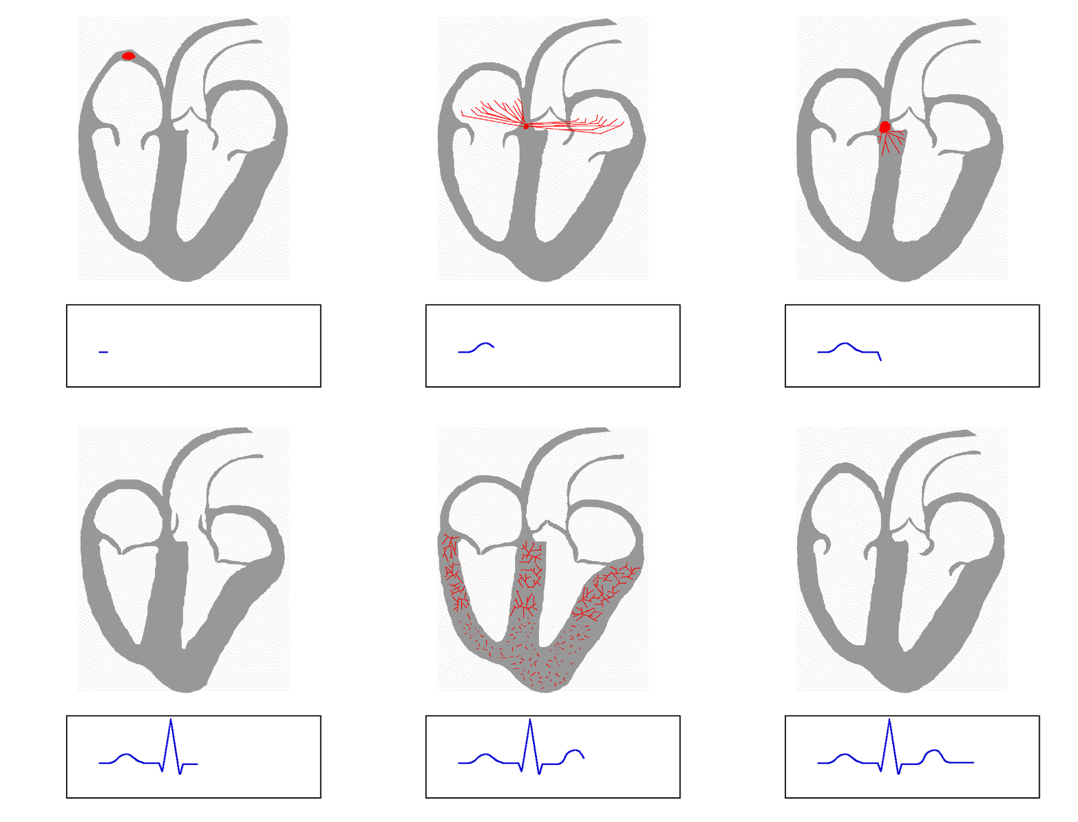
Blood pours into the right atrium from the vena cava and the left atrium from the pulmonary vein.The SA node fires a signal representing a P wave on the ECG. A this point the atria contract, ejecting their blood into the ventricles.As the impulse travels to the AV node, the PR segment shows the pause in electrical activity. This pause allows the ventricles to fill to capacity.The interventricular septum then depolarizes, and then the rest of the ventricular myocardium. We see this as a QRS complex.The blood ejects out of the ventricles. The Ventricular myocardium then recharges which is displayed as a T wave.

The Electrocardiogram - Part III
Adam Thompson, EMT-P | 2:17 PM | Cardiology, Education | 1 comments
Remember how I said that a lead has a positive electrode? Well this comes into play when looking at a specific lead. The isoelectric line is your baseline so-to-speak. The isoelectric line has no amplitude. Anything above the isoelectric line is considered a positive deflection and anything below it is considered a negative deflection.
- When deflections are positive, the mean electrical current is heading in the general direction of the positive electrode.
- When deflections are negative, the mean electrical current is heading away from the positive electrode.
In the above description, you can replace the word 'deflection' with the word 'wave'.
- Deflections or Waves on an ECG strip are representations of heart chamber depolarization and/or replorization (for definitions see basic cardio 2).
Below is an image from Wikipedia which illustrates what I just said:

Take a look at the ECG strip below. Lets say that this is lead II. The yellow circles and lines point out all the positive deflections. The white circle depicts the single negative deflection and the blue lines in the magnified portion are pointing to the isoelectric line.
The isoelectric line is normally at the beginning and end of all waveforms.

EMS Garage Special Edition: How to Improve Survival from Sudden Cardiac Arrest Episode 48
Rogue Medic | 12:30 AM | Cardiocerebral resuscitation, Critical Judgment, Heresy, Rogue Medic | 5 comments
.jpg)
I occasionally appear on EMS Garage. I had to work and missed this episode.[1] I guess that a lot of people will be glad. Just listening to the first few minutes got me started on this post.
First, Mickey S. Eisenberg, MD is the special guest. He has just written a book called Resuscitate!: How Your Community Can Improve Survival from Sudden Cardiac Arrest.[2] He is an excellent person to talk with about this topic. He is one of the people everyone recognizes as an expert. Certainly, I cannot disagree with him, but I do.
Greg Friese is explaining about one of the ideas from the book. That cardiac arrest survival is the best way to determine the quality of a system. I agree that cardiac arrest survival is important, but since cardiac arrest has only been shown to be improved by BLS treatments (compressions and rapid defibrillation - both of which used to be physician-only treatments), it is a mistake to think that this tells you a lot about the quality of an ALS system.
ALS has been shown to worsen the outcome of cardiac arrest, by interfering with good chest compressions, but no ALS treatment has been shown to improve outcome from cardiac arrest. I might even turn that around and say that cardiac arrest outcome may be improved by no ALS.
This may not be entirely true,[3] but it is not unreasonable. There is evidence to show that rapid ALS leads to worse outcomes.[4] There is also evidence to show that ALS leads to worse outcomes.[5]
I do not see evidence that ALS is important in resuscitation. Therefore, how can cardiac resuscitation be an effective measure of the effectiveness of an ALS service?
Cardiac arrest represents about one percent of the EMS calls in any given community, but the management of this one percent encapsulates everything good and bad about a communities EMS system.[6]
In discussing this, Jamie Davis (I think) comments that resuscitation rates are the easiest metric to quantify. I agree that this is easy to quantify. Being easy to quantify and being important do not necessarily go together. The story of the drunk searching for his keys under the streetlight is relevant. He lost his keys elsewhere, but he is looking where the light is better. It will not improve his ability to find his keys, but he will feel better while he is looking. We should not be imitating a drunk, who cannot find his keys. For all we know, they could still be in his pocket, or the bartender might have taken them.
Now that I got that rant out, the rest of the show was excellent. Everybody asked good questions. A lot was covered. The only complaint I have about the rest of the show is that it was too short. There is so much to cover that much, much more than an hour needs to be devoted to this. If I had been involved, the show might have gotten bogged down on the topic I just covered, and never covered some of the much more interesting material that they did cover.
I will write several posts about the many wonderful, positive points in the show.
Footnotes:
^ 1 EMS Garage Special Edition: How to Improve Survival from Sudden Cardiac Arrest Episode 48
EMS Garage
Links to broadcast and downloads
^ 2 Resuscitate!: How Your Community Can Improve Survival from Sudden Cardiac Arrest
By Mickey S. Eisenberg, MD
Amazon.com link with a good video review by Greg Friese.
^ 3 Impact of advanced cardiac life support-skilled paramedics on survival from out-of-hospital cardiac arrest in a statewide emergency medical service.
Woodall J, McCarthy M, Johnston T, Tippett V, Bonham R.
Emerg Med J. 2007 Feb;24(2):134-8.
PMID: 17251628 [PubMed - indexed for MEDLINE]
Conclusions: Highly trained ACLS-skilled paramedics provide added survival benefit in EMS systems not optimised for early defibrillation. The reasons for this benefit are multifactorial, but may be the result of greater skill level and more informed use of the full range of prehospital interventions.
My highlighting, but that may be all that is necessary to explain the benefit. The big question is, Where are the studies showing a benefit from prehospital ALS in cardiac arrest? We can theorize endlessly about potential benefits, but where is the evidence of benefit. It is silly to theorize about the reason for a benefit, when we do not even have evidence that the benefit exists.
Maybe we should be optimizing these systems for early defibrillation or look at systems that have already done this.
^ 4 Cardiac Arrest Survival Rates Depend on Paramedic Experience
Michael R Sayre, Al Hallstrom, Thomas D Rea, Lois Van Ottingham, Lynn J White, James Christenson, Vince N Mosesso, Andy R Anton, Michele Olsufka, Sarah Pennington, Stephen Yahn, James Husar, Leonard A Cobb.
Academic Emergency Medicine; Volume 13 Issue s5; May 2006; pages S55 - S56; abstract number 121
The abstract is available here.
^ 5 Interruptions in Cardiopulmonary Resuscitation From Paramedic Endotracheal Intubation
Henry E. Wang, MD, MS
Scott J. Simeone, BS, NREMT-P
Matthew D. Weaver, BS, NREMT-P
Clifton W. Callaway, MD, PhD
Presented at the Society for Academic Emergency Medicine annual meeting, May 2008, Washington, DC.
Department of Emergency Medicine, University of Pittsburgh, Pittsburgh, PA
The abstract is available here.
^ 6 Resuscitate!: How Your Community Can Improve Survival from Sudden Cardiac Arrest
The Big Picture
Page 18.
Same as footnote [2].
.