Showing posts with label Education. Show all posts

Learn It: Angioedema

Angioedema


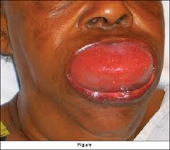
Sometimes referred to as Quinke's Edema, angioedema is that swelling we see that is most apparent around the mucosal areas of the face.  Consider Hives as swelling on the surface of the skin, and angioedema as swelling beneath the skin.  


The most common cause of this type of swelling without the presence of Hives is hypersensitivity to ACE inhibitors.  

ACE = Angiotensin converting enzyme.  This converts angiotensin one into angiotensin two.  

ACE inhibitors block ACE.

Bradykinin is a peptide that has a role with all forms of angioedema.  It is a potent vasodilator that increases permeability and allows the accumulation of fluid within the interstitial space.  

ACE is one of the main ways that bradykinin is degraded.  So when we inhibit the production of ACE, we are then inhibiting the degradation of bradykinin.  We then have this run away peptide and subsequent swelling.  

Many patients that suddenly present with severe angioedema have been taking ACE inhibitors, such as lisinopril, for a long period of time.  They may have never had any issues before, but out of no where have this severe reaction.  This type of reaction is most common in the African-American population, but may occur in anyone.  

There are other types of angioedema, including the traditional allergic reaction.  Those are more well known and prepared for.  


Treatment

As you can see from the pictures above, swelling may be within the oropharynx.  This can cause an airway obstruction, and aggressive airway management should be advocated.  

This patients may be obtunded and snoring as you enter the scene.  They have been confused for diabetics, or acute coronary syndrome patients due to their initial impression.  

It is common for these patients to undergo cricothyrotomy due to complete glottic obstruction.  Moving quickly is imperative to prevent severe hypoxia and cardiorespiratory arrest.

The usual drugs used for anaphylactic reactions are indicated.

- Epinephrine to reduce the vasodilation.  
- Crticosteroids & antihistamines.  


So the next time you run on a patient that is presenting with swelling in the absence of hives, think angioedema, and act fast!


EMS Educast Episode 67


Greg Friese from EMS Educast invited me to guest cohost on episode 67.  On the show was David Page from the St. Paul EMS Academy.

Make sure to go check it out.

Thanks for stopping by,

Adam Thompson, EMT-P

#CoEMS: CPR Effectiveness

Chronicles of EMS, A Seat at the Table takes on CPR effectiveness.  The Las Vegas video that they mention can be found below as well.  Keep up the good work Justin and Mark!

Side note - ILCOR, The International Liaison Committee On Resuscitation has not found any supporting evidence for the Autopulse.  They are the ones whom do the research for AHA.  Also, transporting patients without a pulse should be re-looked at by any agency performing this practice.  The initial treatment at the ER will not differ from the treatment we provide at the scene per ACLS guidelines.  Why not give the patient the best chance possible.  If they don't get a pulse back on scene, it is probably never going to come back--that's just the facts.


Man vs. Machine

Product Review: AHA's Rapid STEMI ID


I was asked a few months ago to review the American Heart Association's STEMI recognition educational software.  This software was created in response to a deficit found during Mission:Lifeline.

Here is the review I gave word for word:


           First, I would like to say that it is not advocated to teach 12-lead ECGs in a STEMI vs. not a STEMI manor.  While the intentions seem good, there is much more to learn regarding 12-lead ECG interpretation that this type of course does not cover.  There is an overwhelming need for a more comprehensive resource for prehospital care providers, and other emergency medical personnel, for that matter. 
            It is my belief that the ACLS course should provide the base of 12-lead ECG knowledge while an online course could be adequate for continuing education.  Initial training could include the six-step method of ECG interpretation.

Six-Step Method:
  1. Rate & Rhythm
  2. Axis determination
  3. Complex, wave, and segment durations
  4. Morphology
  5. STE-Mimics
  6. Ischemia, Injury, Infarct

This method could be taught extensively while breaking down each part to include the physiology of abnormalities.  The Learn: Rapid STEMI ID course is a good start, but is at an educational level below that of what prehospital care providers should be at.  It is also not adequate enough to be part of the curriculum within a paramedic program.  I don’t feel an initial education in 12-lead ECG interpretation should be from an online course because of the inability to ask questions.
Learn: Rapid STEMI ID


Pros:
-       The interactive software is top notch.
-       The graphics used for the cardiology portion are very nice
-       For the most part the cardiology review is very factual
-       There is a good amount of ECG cases
-       The cost seems to be about right for this type of course

Cons:
-       This course is designed only for STEMI recognition, not 12-lead interpretation.  While an important part of 12-lead interpretation, it is not the only ailment that can be determined.
-       With the absence of any presentation outside of STEMI comes the lack of education regarding axis shift, rate changes, pacemaker changes, bundle branch blocks, electrolyte imbalances, etc.  It is possible to create this course in conjunction with more comprehensive resources.  For example, using the six-step method, this course would include steps five and six.
-       The cardiology review is good, but it should also be explained that some patients have slight differences in their coronary arteries (i.e. stenosis, dominant RCA, dominant Cx).
-       During the explanation of electrode application, there should be information about commonly misplaced electrodes and the need to remove all clothing on the patient from the waste up. 
-       There is no list of indications for 12-lead ECG interpretation.  While there is a good explanation of typical and atypical ACS symptoms, ACS symptoms are not the lone reason to acquire a 12-lead ECG.  Some research has shown that paramedics have not performed ECGs on nearly half of patients that present with STEMI at emergency departments. 
-       No information was provided about UA/NSTEMI, informing the learner that it is possible that the patient is suffering from an AMI in the absence of ST-Elevation.
-       It is explained how to identify ST-Elevation, Q-waves, and Hyperacute T-waves, but there is no explanation of the physiology.  A better understanding of the reasoning behind pathological changes will improve the overall efficiency of 12-lead ECG interpretation.   
-       There should be more information regarding posterior wall MI changes (i.e. reciprocal changes in septal leads, R/S ratio >1).
-       One very easy way to determine the J-point’s location is to identify the J-point in a lead above or below the lead in question.
-       Upward concavity is not a conclusive finding with early repolarization.  While the “smiley face method” is a good way to get an idea of the ST morphology, it is not the only way to differentiate early repolarization from STEMI.  Notched J-points, and mean R-wave amplitude in V2-V4 greater than 5 mm are both indicative of benign early repolarization.
-       The information on the STE-Mimics that where covered was good.  However, there is a lot more that could be provided. 
o      LVH was covered, but not RVH
o      Pericarditis symptomology is the easiest way to differentiate it from STEMI.
o      Hyperkalemia vs. Hyperactute T-waves
o      Bundle branch blocks
o      Brugada Syndrome
-       Contiguous leads should have been explained better V1 & V6 are not contiguous. 
-       The differentiation between Septal, Anterior, and Low Lateral should be made.  V1-V6 are not all considered anterior. 
-       The depth of Q-waves was not covered pathological vs. physiological.  Width was appropriately taught, but not depth.
-       It should be made known that MI is not the most common cause of ST-Elevation.

Overall, I am pleased to see that the AHA is making an attempt at 12-lead ECG education.  I am a big fan of the AHA and its use of evidence-based medicine.  In fact, there is plenty of evidence supporting ECG findings, and a need for better interpretation.  Here is an example of something that might be missed out on if this course was the base of the responding paramedic’s ECG knowledge; patient with syncope who has long QT syndrome or Brugada syndrome.  This patient may never even have a 12-lead ECG obtained even though that the two conditions described can both be lethal.  I am optimistic about the possibilities to come.  


Advocating Airway Education

In the popular and acclaimed JEMS article Experts Debate Paramedic Intubation, there were a few key points made that I would like to elaborate on, as well as provide some of my own insight from the research I have come across.


Key Point 1

Endotracheal Intubation has been best performed by those who maintain experience and those whom utilize Rapid Sequence Induction/Intubation.

Experience should be maintained in a number of manors:
  • Operating room rotations
  • Mannequin scenarios (without the dummy supine on a table)
  • Cadavers if possible
Rapid Sequence Induction is when one of many combinations of sedatives and paralytics are used to facilitate endotracheal intubation.  This is a high risk procedure with many possible complications.  It requires more education, and practice.
Dr. Bledsoe: Do you feel there’s a role for RSI in the prehospital setting? Dr. Wayne, I know your program has decades of success with RSI. What do you think?
Dr. Wayne: Although there are no nationally defined indications for the use of RSI in the field, we at Whatcom Medic One believe that RSI is indicated for any patient in whom there’s a need to control an “uncontrolled” airway. This may include depressed GCS score, excess secretions, hypoxia that may be correctable, ventilatory fatigue or central nervous system depression with or without secondary respiratory depression.
Dr. Tan: I believe there is, but it must be in the right context with requisite oversight and extraordinary training. I oversee more than 100 paramedics in my system, yet only 10 of them have RSI privileges. They’re required to obtain critical care certification, attend ongoing training sessions with me every 12 weeks, attend annual specialized training courses and undergo 100% audits of their critical care trips. It’s a strenuous and time-consuming process but one that can’t be overemphasized given the complexity and danger inherent to RSI. I certainly don’t believe RSI should be a “routine” part of any standing orders, as there is nothing routine about it.
Dr. Wang: I think RSI should be restricted to the aeromedical setting for use by critical care flight nurses and/or flight medics for the reasons I’ve previously detailed. I really challenge those medical directors who currently allow RSI and promote its use in other systems. Although I applaud their efforts and attention to quality improvement and training, they still equate successful intubation with a positive outcome. As Dr. Eckstein said, in the absence of prospective RCTs, we can’t assume that prehospital RSI has actually improved outcomes for our patients.
Dr. Eckstein: RSI is potentially useful where paramedics have exceptional skill, training and medical oversight. Unfortunately, this is a tiny fraction of EMS agencies. If we replaced the “I” (intubation) with “A” (airway—Combitube, King, etc.), this might relieve much of the angst over prehospital RSI.


Key Point 2
Airway Management ≠ Endotracheal Intubation (ETI)

What I mean by that, is that just because a patient's airway requires management, it does not mean that ETI is the only option.

Questions to ask:
  • Is there a risk for aspiration?
  • Is the patient ventilating on their own?
  • Is the patient oxygenating on their own?
  • Is the patient conscious?
  • How difficult will this ETI attempt be?
  • What is my backup plan?
Other options:
  • Bag-valve mask (possibly with an OPA/NPA)
  • Combi-tube
  • King LT/LTD
  • Laryngeal Mask Airway
Dr. Bledsoe: Are the alternative airway devices (e.g., King LT, etc.) good enough for prehospital airway management?
Mr. Gandy: Yes. The studies have shown that excellent ventilation can be achieved with these devices.

Key Point 3


The #1 way to confirm proper placement of the endotracheal tube in the field is end-tidal CO2 (ETCO2).  If you have ETCO2 available in the field, use it.  


ETCO2 measures the amount of CO2 that is being exhaled by the patient.  This lets us know that the O2 we are putting into the body is being used and exchanged for the CO2 that comes out via pulmonary perfusion.  This exchange occurs in the lungs, which just so happens to be the place that we are attempting to ventilate.


Key Point 4

Anticipate the difficult airway.


Mr. Gandy: The biggest problem is inadequate training and practice in airway evaluation, such as using the Malampatti or Cormack-Lehane criteria; using aids to intubation, such as bougies; the BURP maneuver; alternative laryngoscope techniques, such as the “skyhook” technique; and a good assortment of alternative airway devices, including either GlideScope or AirTraq. Ventilation should be emphasized over intubation, and extensive practice with BVM ventilation should be required.

Malampatti scoring is done by having the patient stick out their tongue.  The difficulty of the proceeding ETI attempt can be gauged by the visibility of the oropharynx.


Don't aim for jewelry!


Cormack-Lehane Citeria is utilized with direct laryngoscopy.  This is done by visualizing the vocal cords and making note of how much of the opening is visible:

  • Grade 1, visualization of the entire laryngeal aperture; 
  • Grade 2, visualization of parts of the laryngeal aperture or the arytenoids; 
  • Grade 3, visualization of only the epiglottis; and 
  • Grade 4, visualization of only the soft palate.

Bougie - This is almost like a super long rigid stylet that is introduced through the vocal cords first.  You then thread the ET tube over it.   


BURP Maneuver - Backward, Upward, Rightward, Pressure of the larynx.


Don't worry if you don't understand the picture above.  It is just a step by step of the BURP maneuver.  Basically you place your fingers on the palpable cricoid ring of the patient.  Push towards their posterior, and slightly towards their right.  This should bring the trachea and it's structures to the best point of view during direct laryngoscopy.


"Skyhook" - I believe Gandy is referring to what my peers and I call the "fish hook" maneuver.  This is reserved for the more hefty patients that may be hard to intubate.

This is a two person procedure.  One person is dedicated to laryngocopy, and the other will direct person 1, visualize the vocal cords, and pass the ET tube.

Person 1 - With Laryngoscope and a Macintosh blade

- Straddle the supine patient
- Hook the blade into the mouth
- Pull back, keeping the blade off of the teeth
- Make adjustments based off person 2's direction

Person 2 - With appropriately sized ET Tube

- Position yourself at patient's head
- Direct person 2 until the vocal cords are visible
- Pass ET tube


I spoke about the Glidescope in my post Video Laryngocopy.  Go check it out.


Key Point 5


It doesn't end after the intubation is accomplished.


Once you've got the tube, you should aim all of your efforts at keeping the tube and ventilating ACCURATELY.  Using a mechanical ventilator after the ET tube is placed provides the ability to set an accurate rate and tidal volume.  If one is not available, ETCO2, and O2 saturation should guide your ventilation rate and tidal volume.  

Place a cervical collar on the patient to limit their movement.  

Make note of the depth,

Monitor diligently. 

It isn't the end of the world if you lose the tube.  It may be the end of your career if you don't realize it.

Please see Post-Intubation Tracheal Stenosis for yet another consideration.


Treating Tachycardia


Treating Tachycardia
By Adam Thompson, EMT-P


Tachycardia simply means a faster heart rate than normal.  With the sinoatrial node, which is the heart's inborn pacemaker, the intrinsic rate is between 60 and 100 beats per minute.  When the rate exceeds 100 beats per minute, tachycardia is present.

When treating tachycardia, it is important to first consider a compensatory cause.  The body tends to use an increased heart rate as a frequent compensatory mechanism when it senses decreased perfusion.  Two of the best dysrhythmics in the EMT and paramedic's tool box are OXYGEN and NORMAL SALINE. Both of these treatments should be attempted prior to using any other medication.  It is not advantageous to eliminate a compensatory tachycardia in a patient who needs it to perfuse.  Locating the cause of the decreased perfusion would be optimal.

Another thing to consider is the patient's hemodynamic stability.  With organized tachycardic rhythms in unstable patients, synchronized cardioversion is indicated.  There seems to be a fear amongst prehospital providers when it comes to shocking people.  The paramedic seems to be much more comfortable giving anti-arhythmic/dysrhythmic medications than they do performing cardioversion.  This is in-fact backwards thinking.  Consider Kelly Grayson's outlook on dysrhythmic drugs--they are selective cardiotoxins.  First off, they are not naturally found in the body.  Second, they metabolize over time and the reaction can be unpredictable.  Thirdly, they are used to counteract cellular depolarization.  Do you know what happens in the absence of cellular depolarization in the myocardium?  Asystole--not a common side effect, but it drives home the point doesn't it?.  Other complications, like high-grade atrioventricular blocks, and long QT syndrome may also occur.  Conversely, synchronized cardioversion doesn't have nearly as many unwanted effects.  It works fast, and goes away.  The medication you should be considering, is some sort of sedative or benzodiazapine prior to cardioversion.

Next, after determining the patient's hemodynamic stability, the width of the QRS should be considered. If the patient is stable, and they are in a sustained tachycardia, dysrhythmic medications can be considered.  It is important to determine the width of the QRS, because medications like Cardizem (diltiazem), or Adenocard (adenosine) that may be administered to narrow complex rhythms, can effectively KILL people with wide QRS rhythms.  Notice that there is not a 'ventricular tachycardia' algorithm?  It states 'Wide QRS', and lists 'uncertain rhythm' below.  This is an important concept.  If it is wide, and you are uncertain of the origin, it is ventricular tachycardia until conclusively proven otherwise.  Another reason that it is a WCT guideline and not a ventricular tachycardia guideline is because of conditions like WPW (wolff parkinson white syndrome).  With WPW, a delta wave may be present causing widening of the QRS complex.  This is important because adenosine, and Cardizem should not be administered to patients with WPW.  There is controversy regarding whether Amiodarone is safe with WPW, but as of now the American Heart Association considers it a safe option.

A wide QRS complex is considered greater than 120 ms or 0.12 seconds or 3 small boxes.


Points to remember:

  • O2 & fluids for compensatory tachycardia
  •  Synchronized cardioversion is the SAFER option
  • If QRS is wide treat as V-tach
Note: Torsades de Pointes should not be treated with Amiodarone.  This can cause lengthening of the QT interval, and subsequently a worse arrhythmia.  


Brugada Criteria.  This should only be used to confirm ventricular origin.  Not to rule it out.  

View more documents from Adam Thompson.

Videos: Acute Coronary Syndrome

Some stuff for you visual learners, enjoy.


This one ends abruptly, but makes a point that I like to stress, DIAPHORESIS is BAD!

Sorry for this one, but I thought it was funny


In the following video, the presenter has used some of Tom B's teaching aids to teach Sgarbossa's criteria.  I pronounce it with the 'S' by the way.  

Also check these out - video , & video

Drug Profile: Ketamine

Ketamine 

By Adam Thompson, EMT-P


Since my beginning in the world of prehospital medicine, which wasn't too long ago, I have heard more and more about a particular drug.  I have attended critical care and emergency medicine conferences and the same has been true.  Ketamine seems to have become a favorite amongst many physicians.  I have never administered the drug myself, but it has made its way into some prehospital standing orders, and with reason.  


Ketamine Hydrochloride
Non-barbiturate anesthetic 

Mechanism of action:
  • Creates a state of dissociation from reality.  
  • Catecholamines (epinephrine & norepinephrine) are released.  
  • Spinal reflexes are reduced.


Indications:
  • Indicated for anesthesia when cardiovascular depression must be avoided.  
  • A commonly used induction agent to facilitate endotracheal intubation.

Contraindications:
  • Hypersensitivity to Ketamine
  • Increased intracranial pressure
  • Hypertension
  • Aneurysm
  • Thyrotoxicosis (hyperthyroid)
  • Congestive heart failure
  • Angina
  • Psychotic disorders
  • Pregnancy
So there are a lot of contraindications that happen to be conditions that we see commonly.  But think about what they are, and what the side effects are of the other drugs that we administer for similar indications.  Almost every other medication decreases blood pressure.  Ketamine actually increases it, so it has its place.  

Side Effects:
  • Hallucinations
  • Vivid dreams
  • Hypertension
  • Increased cardiac output
  • Tachycardia
  • Paradoxical direct myocardial depression
  • Increased ICP
  • Tonic-clonic movements

Overdose:

      With high doses or rapid administration, respiratory depression may occur.


Dosage:
  • Oral: Pediatric 6 - 10 mg/kg
  • Intramuscular: 3 - 8 mg/kg
  • Intravenous: Pediatric 0.5 - 2 mg/kg, Adult 1 - 4.5 mg/kg
Onset is about 1-2 minutes if given I.V., and 3-8 minutes if given I.M.


Some research:

The effect of combined treatment with morphine sulphate and low-dose ketamine in a prehospital setting [1] 
Conclusion 
We conclude that morphine sulphate with addition of small doses of ketamine provide adequate pain relief in patients with bone fractures, with an increase in systolic blood pressure, but without significant side effects.
Anesthesia in prehospital emergencies and in the emergency department. [2]
Abstract
PURPOSE OF REVIEW: Recently, notable progress has been made in the field of anesthesia drugs and airway management. RECENT FINDINGS: Anesthesia in prehospital emergencies and in the emergency department is reviewed and guidelines are discussed. SUMMARY: Preoxygenation should be performed with high-flow oxygen delivered through a tight-fitting face mask with a reservoir. Ketamine may be the induction agent of choice in hemodynamically unstable patients. The rocuronium antagonist sugammadex may have the potential to make rocuronium a first-line neuromuscular blocking agent in emergency induction. Experienced healthcare providers may consider prehospital anesthesia induction. Moderately experienced healthcare providers should optimize oxygenation, hasten hospital transfer and only try to intubate a patient whose life is threatened. When intubation fails twice, ventilation should be performed with an alternative supraglottic airway or a bag-valve-mask device. Lesser experienced healthcare providers should completely refrain from intubation, optimize oxygenation, hasten hospital transfer and ventilate patients only in life-threatening circumstances with a supraglottic airway or a bag-valve-mask device. Senior help should be sought early. In a 'cannot ventilate-cannot intubate' situation, a supraglottic airway should be employed and, if ventilation is still unsuccessful, a surgical airway should be performed. Capnography should be used in every ventilated patient. Clinical practice is essential to retain anesthesia and airway management skills.
Pre-hospital use of ketamine for analgesia and procedural sedation [3]
Abstract
The safe delivery of adequate analgesia and appropriate sedation is a priority in prehospital care. The use of ketamine is described for analgesia and sedation in 1030 trauma patients in a physician-led prehospital trauma service. Ketamine was mainly used in awake non-trapped patients with blunt trauma for procedural sedation and analgesia. Detailed database searches did not demonstrate loss of airway, oxygen desaturation or clinically significant emergence reactions after ketamine administration. Ketamine is relatively safe when used by physicians in prehospital trauma care.
Ketamine for prehospital use: new look at an old drug. [4]
Abstract
INTRODUCTION: Ketamine has been used extensively for analgesia and anesthesia in many situations, including disaster surgery where extra personnel and advanced monitoring are not available. There are many features of ketamine that seem to make it an ideal drug for prehospital use. The reported use of ketamine in the prehospital environment is limited, however. The purpose of this study is to review the experience in the use of ketamine in a regional air ambulance service and suggest indications for its use in the prehospital setting. METHODS: This was a retrospective study of all patients transported by a regional aeromedical program. Patients were included in this study if the crew had used ketamine at any time during the flight. Data regarding the transport collected included patient age, type of transport, indications for ketamine use, and adverse reactions. RESULTS: During the period studied, ketamine was used in 40 patients. The age range was 2 months to 75 years. The indications and situations requiring use were varied and included both trauma and medical patients. Hypotension with need for analgesia, agitation or combativeness and intact airway, or pain unresponsive to narcotic medications were the most common indications for use. Ketamine was used both intravenous and intramuscular, even without intravenous access. There were no adverse reactions. CONCLUSIONS: Ketamine is an ideal drug for use in many prehospital situations. Our experience suggests that it is safe, effective, and may be more appropriate than drugs currently used by prehospital providers.


When All You Have is a Hammer, Every Problem Looks Like... Lasix?

I posted this article over at http://www.lifeunderthelights.com/ a few days ago and I thought it would good for here as well. I'm sorry that I've only rarely posted here, but I read this blog quite often and recommend it to all of my peers. Enjoy

-----------------------------------------------

A few years ago I responded to a structure fire on the main engine out of my station. The fire was at a house that had been converted to a dog kennel and grooming shop just a few blocks away from the firehouse and was a short response time. It was a light-staffing day and we responded as a three person engine company. As the senior firefighter I was the acting company officer and my new girlfriend at the time, who just happens to be my wife now, was the backseat firefighter. Get ready for the “Awwww” moment… it was our first fire “as a couple”. There was a number of cool things that came out of the fire, but one of them was the fact that Gina grabbed *my* maul.

On our main engine, there’s an 8-pound maul (big hammer) that I grab as my tool of choice every time I jump off the truck for a fire. It just tucks so neatly in my SCBA’s belt and is so compact yet handy that I make a beeline for it every time. This time, Gina had taken it, so I grabbed a pick-head axe.

It’s amazing when I have my maul how every access problem looks like something that I can solve by whacking it with a hammer of some sort. On this fire, I learned that when one has an axe, every problem looks like it can be solved by some sort of chopping.

Moral of the story, Gina and I entered the structure, saved the pooches, and stopped the fire in its trucks with minimal damage. There’s actually a hilarious video that I believe is still on our department’s web site that I’d let you see if I didn’t hide the name of the department(s) I work for due to reasons of wanting to remain employed.

And, like a lot of posts I write, I told you that so I could tell you this about an EMS call I responded to an indeterminate amount of time ago. I have the honor and privilege to be the senior medic on most shifts I work and I precept a lot of students on the ambulance. This shift was no different and this 0-dark-30 call illustrates a point that I’d like to explain to you.

For this call, the primary ambulance out of our station responded because they were on the way back from another call and my partner and I responded in our ambulance because we were up on the alternating call rotation. They arrived at the poorly-accessible apartment complex a few minutes before we did and made first patient contact. As it turns out, the middle age patient had ran out of his/her prescription Lasix (a potent diuretic, or water pill) a week or so prior to the call and had been retaining a great deal of excess bodily fluid. The patient’s legs were markedly and grossly swollen and weeping fluid out of fluid filled blisters. The Patient called us because he/she could no longer stand the pain of the cellulitis (infection) that had developed. The patient had no respiratory compromise, his/her lungs were clear, and he/she really had no other complaints. The patient had an extensive medical history of organ failure and disease. He/she was fully alert and oriented, and was able to assist us as we simply picked him/her up and carried him/her to the cot.

As we were loading the patient up in the ambulance and I was about to get into the back to continue my assessment and treatment of the patient, the EMT from the other ambulance who happens to be an almost-done Paramedic student told me, “So those legs are the worst I’ve ever seen fluid wise, you’re going to push some lasix on this one”. I mumbled something and got into the truck. I was tired and wasn’t really able to form complete sentences at the time due to sleep deprivation. I continued my assessment where I found that the frail patient had a blood pressure in the 70 systolic range (Low!) and that in addition to retaining fluid in his/her legs, he/she was also retaining fluid in his/her abdomen and was probably in need of a paracentesis. I managed the patient with a (beautifully executed, I must say) IV stick into an impossibly small and crooked vein, and gave just enough fluid to bring his/her BP up a bit without adding to his/her fluid overload all that much. I put the Pt on oxygen and a cardiac monitor, which revealed a normal sinus rhythm without ectopy and obtained a 12-lead EKG as well, which was not indicative of any acute problems. The patient stated that his/her pain was managed by padding and positioning of his/her swollen legs and even though he/she complained of no breathing problems, I put him/her on a bit of oxygen via nasal cannula.

The transport was uneventful, although his/her blood pressure never did come up. The ER later diagnosed the Pt with complete liver failure and toxicity.

But the interesting part of the story is this, when I got back the medic student asked me about giving IV lasix to the patient, as we carry that in our medication stock and have it available as an emergency diuretic for patients in congestive heart failure and/or fluid overload with pulmonary edema and respiratory compromise. He was almost taken aback when I said that I didn’t give any.

I asked him if he did a full assessment. He said that he had tried… but that he didn’t have enough time before I arrived and we took the patient out to the ambulance. I gave him my assessment findings and the news of the very low blood pressure. He said that he agreed with me on not giving the lasix with the markedly low blood pressure but was curious when I explained that it wasn’t the reason I didn’t give the medication.

We in EMS, and especially new providers carry our own hammers… our treatments and medications that we’re able to give in the field. Medics that use these treatments more often are called “aggressive” and it is a badge of honor. In fact, in some cases, aggressive field treatment is indeed warranted and improves patient outcomes. However, in a lot of cases it is not indicated and patients benefit from what we don’t do more so than from what we could have done.

This patient didn’t have any respiratory compromise and while he/she obviously could have benefited from the dieresis or removal of the excess fluid, she didn’t meet the criteria for emergent field administration of lasix, which is respiratory compromise from pulmonary edema. I made the decision to let the physician evaluate the patient and determine the best treatment path that would fit in with the patient’s ultimate plan of care. I didn’t believe that the patient would ultimately benefit from my administration of lasix twenty minutes earlier than the ER could have done it if the physician so chose.

Every treatment we administer must be given with a full assessment of the risks and benefits to the patient for doing so. Every EMS person should familiarize themselves with the long-term care paths of the conditions we treat and try to maximize the long-term benefit to the patient with the acute and short-term care we give. Not every problem is “a nail” and sometimes the hammers we carry aren’t the best ultimate solution for excellent patient care. Remembering how we as EMS people fit into the grand scheme of the overall healthcare system and in the ultimate care paths of our patients will help us all to do what we’re supposed to do, which is to provide excellent and appropriate patient care.

It is also of note, I guess, that Gina rarely steals my maul anymore. Now that we’re married… I “give it freely” to her.. What’s mine is her’s, as they say.

--------------------------------------------
 
The original post has some pictures of the fire and of the doggies that my wife and I saved on our first "Fire Date" - It can be seen HERE

12-Lead Differential Diagnosis: Syncope


12-Lead Differential Diagnosis: Syncope
By Adam Thompson, EMT-P


There are many causes of syncope. Syncope is the medical term for fainting. Most of us are pretty familiar with the common vasovagal cause. Fortunately syncope is often self-correcting; the patient hypo-perfuses, they blackout and fall, their body perfuses better, they wake up. This post will be dedicated to 12-lead presentations that may indicate causes of syncope.

I have decided to use some exerts from my favorite emergency physician/presenter, Dr. Amal Mattu.  Dr. Mattu has spoke and written on these topics multiple times.  


Bradycardia

This first one is easy, and you wouldn't even need a 12-lead to determine it's malignancy.


What do you notice about the 12-lead above? How about the rate? This is an example of bradycardia. Bradycardia may cause hypo-perfusion, leading to syncope. This would classify them as symptomatic, and they may require treatment.

*It is important to remember that there are many tachycardic arrhythmias that could cause syncope as well.   


Acute Myocardial Infarction

An acute myocardial infarction (AMI) is the most common reason we use a 12-lead for diagnostic purposes. An AMI may cause syncope amongst many other signs and symptoms.  Syncope would be an atypical (not usual) presentation for an AMI.  I am not going to elaborate much on this presentation because it requires much teaching for those who are unfamiliar with STEMIs (ST-Elevated Myocardial Infarction). Please head over to the prehospital 12-lead blog for some great education on STEMIs. 


Long QT Syndrome


Long QT Syndrome, or LQTS may lead to arrhythmias that lead to syncope.  This happens due to something called an R on T phenomenon.  The most common arrhythmia due to LQTS is Torsades de Pointes, however monomorphic ventricular tachycardia is possible.  Syncope and/or seizures are common symptoms of the rhythms associated with LQTS.

Torsades de Pointes

From Dr. Amal Mattu:


Prolonged QT-Interval  
Prolonged QT-interval predisposes to torsades de pointes
One of the key “can’t miss” diagnoses associated with syncope
Perhaps a more common cause of syncope and sudden death than previously recognized?

Causes of QT-prolongation
Electrolyte abnormalities (hypokalemia, hypomagnesemia, hypocalcemia)
Sodium channel blocking medications (many!)
Includes Type IA medications, anticholinergics, cocaine, many antipsychotics,  some antibiotics
Acute myocardial ischemia (usually associated with inverted T-waves)
CNS lesions, e.g. intracerebral hemorrhage (often associated with giant inverted T-waves)
Hypothermia
Congenital

How long is too long?
QT-interval will vary based on patient’s heart rate
Measure QT from beginning of the QRS complex to the end of the T-wave, and average over 3-5 beats
“Corrected” QT-interval (Bazett formula): QTc = QT/(RR)
QTc is considered prolonged when > 450 msec in men and > 460 msec in women and children
Major risk occurs in patients with QTc > 500 msec

Treatment
Search for and correct underlying cause (e.g. correct electrolyte abnormalities, discontinue responsible medications, etc.)
Congenital or idiopathic causes: beta-blocking medications attenuate adrenergic-mediated trigger mechanisms 
Treatment of torsades de pointes: cardioversion/defibrillation, magnesium if relatively stable (e.g. intermittent torsades): 2 grams IV over 2-3 minutes followed by infusion
Overdrive pacing? Isoproterenol? Atropine? These are listed as possible treatments in acquired (not for congenital) cases, but rarely needed
Post-conversion treatment with magnesium, not lidocaine/amiodarone/ procainamide! (unlike other forms of ventricular tachycardia); for congenital cases, add beta-blocking medications


Click here or here to learn about Long QT Syndrome


Brugada Syndrome


ECG example of Brugada Syndrome


Brugada syndrome is becoming more and more well known recently.  It is associated with specific ECG changes and an increased risk for sudden cardiac arrest.  Only known cause of Sudden Unexpected Death Syndrome (SUDS)--according to Wikipedia.  The ECG changes associated with Brugada Syndrome are most visible in V1, and V2.  Non-STEMI-like ST-elevation that cannot be explained by another pathological cause (ie. LVH, LBBB, BER) may be Brugada Syndrome.  These individuals may be otherwise very healthy and/or young.

Some features of the different types of Brugada Syndrome include:


  •  a RBBB pattern in V1 without terminal S-waves in lead I and V6.  
  • A saddleback ST-elevation pattern (type 2 below)
  • Coved J-point elevation in V1, V2, V3 that gradually slopes down (type 1 below)


From Dr. Amal Mattu:

Brugada Syndrome  
More common cause of sudden death than previously recognized
May be responsible for up to 20% of sudden deaths in individuals without structural heart disease
Responsible for 4-5% of all sudden deaths
Incidence varies in different populations (some genetic factors involved)
Most common in young males (< 50 yo.)
First onset of symptoms approximately 40 yo.
Mortality approximately 10% per year if not treated with an internal cardioverter-defibrillator (ICD), regardless of whether or not antiarrhythmics are used  
Syndrome characterized by
ECG abnormalities in leads V1 – V3
Polymorphic or monomorphic (less common) ventricular tachycardia
Causes syncope if self-terminating
Causes sudden death if persists and not terminated by treatment
Structurally normal heart
Familial occurrence in approximately half of patients  
ECG findings in leads V1 – V3
Right bundle branch block (RBBB) or incomplete RBBB pattern
ST-segment elevation — 2 types
“coved-type” (most common)
“saddle-type”
ECG findings can vary with time depending on the autonomic balance, administration of antiarrhythmic and other drugs affecting channel function, body temperature, and other unknown factors  
Definitive diagnosis can be made with electrophysiologic testing
Challenge with an intravenous class I medication (e.g. ajmaline, procainamide, flecainide)
Will induce increased ST-segment elevation and “coving”
Programmed electrical stimulation of the heart
Can induce ventricular tachycardia  
Treatment
Placement of an ICD is the only effective treatment
Antiarrhythmic drugs (including beta blockers, amiodarone, etc.) ineffective 

Click here or here for more on Brugada Syndrome


Hypertrophic Cardiomyopathy


From Dr. Amal Mattu:


Hypertrophic Cardiomyopathy (AKA IHSS, ASH, HOCM, etc.)

Prevalence — 0.02% – 0.2% of the general population
Genetic factors involved
Hypertrophied but nondilated left ventricle
Thickening is usually asymetric, involving the septum to a greater extent than the free ventricular wall
Cardiomegaly usually not present on chest x-ray
Mortality 3.5% per year
Diagnosis often made only when the patient experiences sudden death
Usually occurs during exertion \
Average age at diagnosis is 30 – 40 yo.
Patients may also experience syncope, angina, palpitations, dyspnea (often associated with exertion)  
ECG findings
Normal in 7% – 15%
Typical abnormalities
Deep narrow Q-waves in the inferior and/or lateral leads – I, aVL, V5-6 (simulates MI, but Q-waves are “too narrow” for MI)
Very specific for this condition
Q-waves in lateral leads are more common than inferior leads, very commonly misdiagnosed as lateral MI
Left atrial enlargement
High left ventricular voltage/left ventricular hypertrophy
Other less common abnormalities
Tall R-wave in lead V1 (simulates posterior MI)
Deep narrow Q-waves in the inferior leads (simulates inferior MI)
Don’t rely on your cardiologists to make the Dx on ECG! Clinical diagnosis
Systolic murmur at apex or LLSB
Murmur increases with valsalva, standing
Murmur decreases with trendelenburg position, isometric exercise, squatting
Definitive diagnosis — doppler echocardiography  
Treatment
Beta blockers, calcium channel blockers to improve LV filling and diastolic \function ]
Amiodarone if ventricular dysrhythmias present 


Unrecognized Killers in Emergency Electrocardiography 
 Amal Mattu, MD 7